Fig. 8.

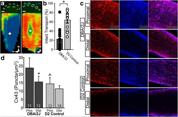
Diminished anterograde transport in a sample of DBA/2 J nerves. a Top Left: Coronal section through superior colliculus of DBA/2 J mouse (between dashed white lines) following intravitreal injection of CTB (green). Bottom Left: Corresponding retinotopic map shows nearly depleted anterograde transport of CTB. Top Right: Coronal section through superior colliculus of D2 control mouse prepared as on left. Bottom Right: Corresponding retinotopic map shows a full complement of anterogradely transported CTB. b Transport of CTB from DBA/2 J eyes was near 20% (22.8 ± 5.5), significantly reduced from D2 control which is near 75% (74.896 ± 4.328) (p = 0.0015). c Confocal micrographs of proximal (left) and distal (right) DBA/2 J (top) and D2 control (bottom) optic nerves. Connexin 43 (Cx43, blue) and GFAP (red) colocalize, and both are elevated in proximal optic nerve. d. Density of Cx43 (puncta/μm2) in proximal segment of DBA/2 J nerves is significantly elevated compared to both distal DBA/2 J (*; p = 0.043) and proximal D2 nerves (#; p = 0.032)
