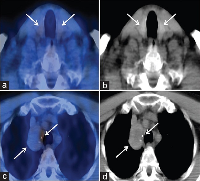
Figure 1.

Fused axial positron emission tomography/computed tomography and low-dose computed tomography images of lower neck (a and b) showing nonhypermetabolic orthotopic bilateral thyroid with heterogeneous computed tomography appearance. Fused axial positron emission tomography/computed tomography and low-dose computed tomography images of the upper chest (c and d) demonstrating nonhypermetabolic right paratracheal mass with central hypodensity and peripheral calcifications. Mild uptake at the medial aspect of the mass represents inflammation at biopsy site
