Figure 3.

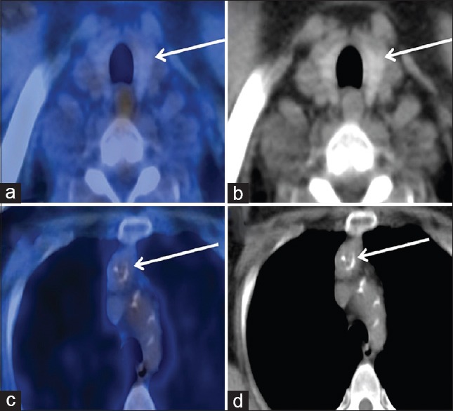
Fused axial positron emission tomography/computed tomography of the neck (a) and noncontrast computed tomography (b) of the neck showing normal appearing thyroid gland with physiological uptake. Fused axial positron emission tomography/computed tomography of the chest (c) and noncontrast computed tomography (d) of the chest showing a mildly active heterogeneous anterior mediastinal mass with calcifications abutting the posterior aspect of the left brachiocephalic vein and aortic arch. This was confirmed as ectopic thyroid on biopsy
