
Figure 7.
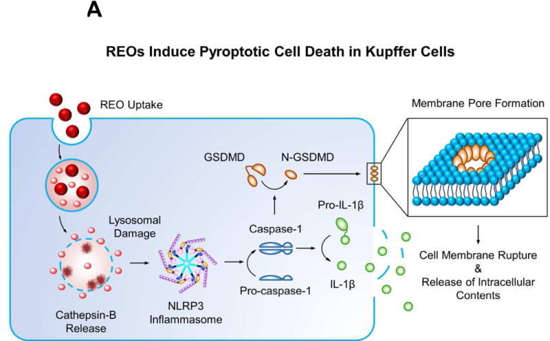
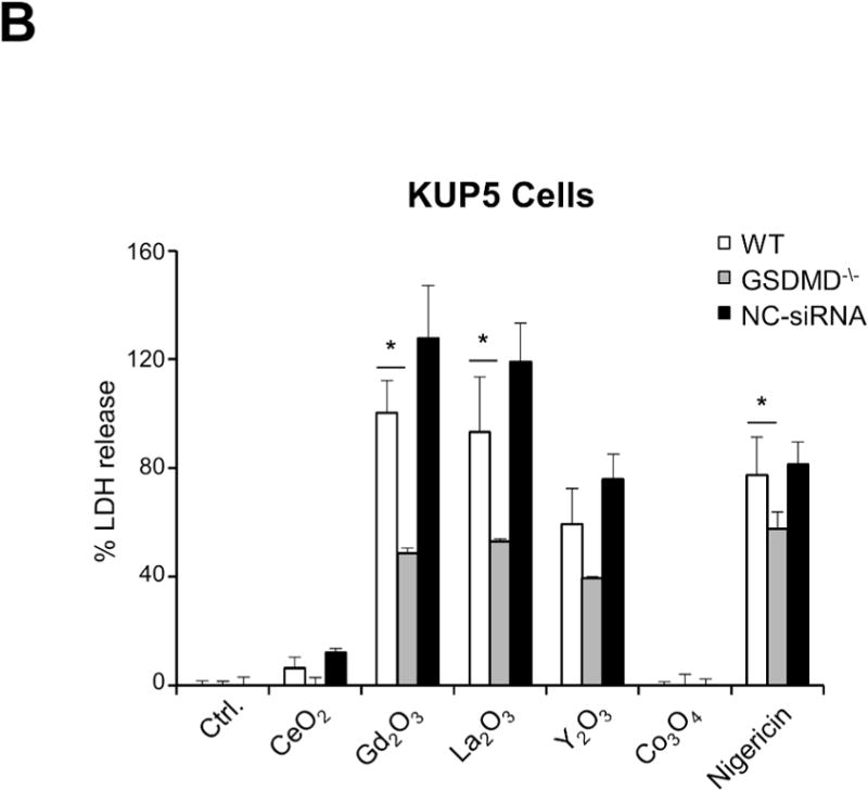
Confirmation that the induction of cell death by REOs represents a pyroptosis process in Kupffer cells. (A) Schematic depicting the possible pathway by which REOs induce pyroptosis. Intralysosomal particle biotransformation leads to damage to the organelle and NLRP3 inflammasome activation. The accompanying activation of caspase 1 leads to the cleavage of pro-IL-1β, as well as gasdermin D (GSDMD), leading to the generation of a N-terminal fragment, N-GSDMD. Oligomerization of N-GSDMD in the cell membrane leads to the formation of pores, cell swelling, release of pro-inflammatory products and ultimately the features of pyroptotic cell death. (B) LDH cellular release to show that siRNA knockdown of GSDMD ameliorates REO-induced cell death in KUP5 cells. LPS-primed (1 μg/mL, 4 h) KUP5 cells were exposed to 200 μg/mL of the chosen MOx nanoparticles for 3 h. Cellular supernatants were collected to assess LDH release by a CytoTox 96® NonRadioactive Cytotoxicity (LDH) Assay kit. Nigericin (10 μM) was used as a positive control. *p<0.05 compared to particle treated wild type KUP5 cells.