
Figure 8.
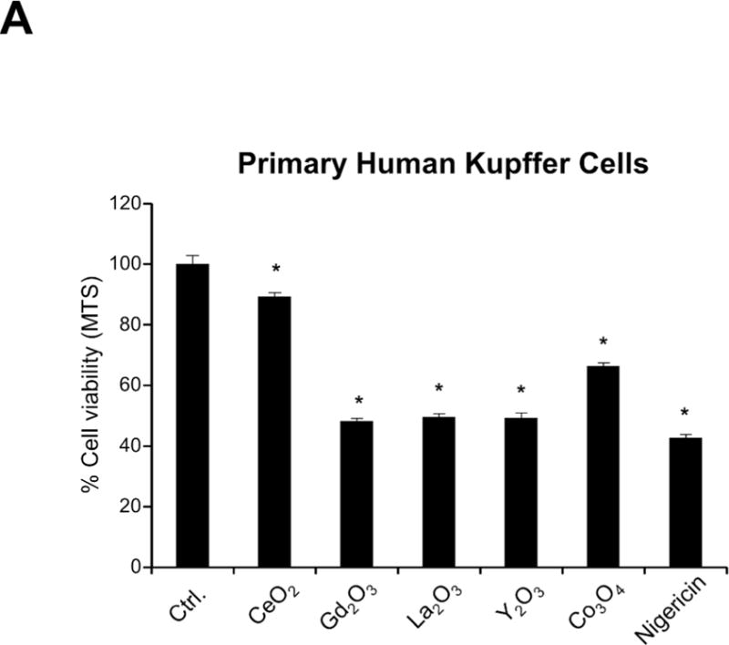
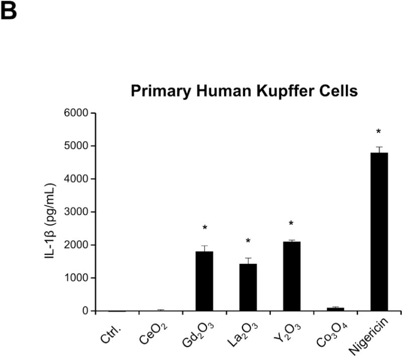
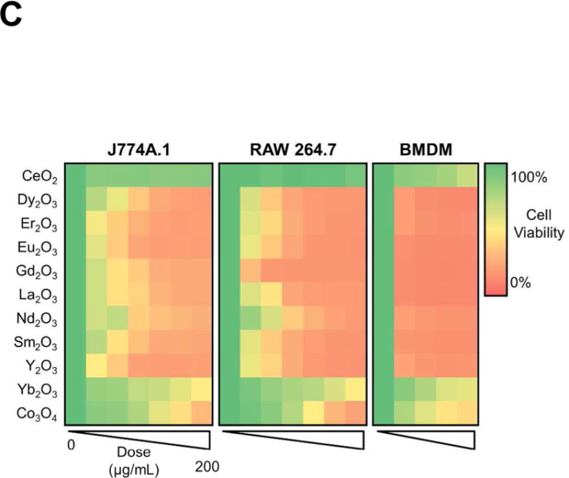
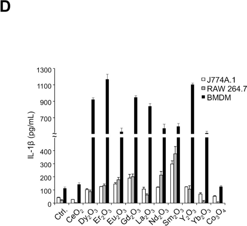
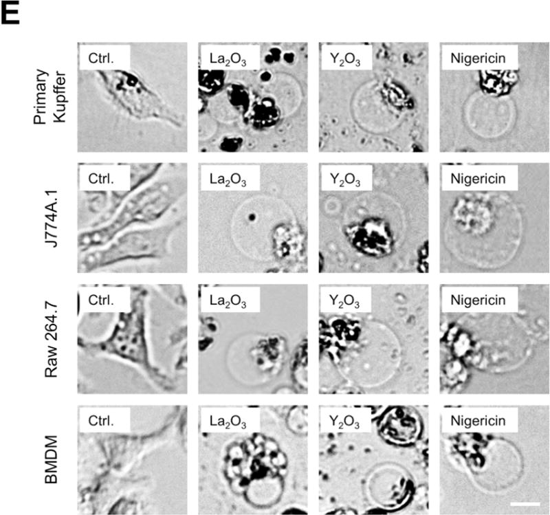
REOs induce pyroptosis in primary human Kupffer cells and other phagocytic cell types. (A) LPS-primed (1 μg/mL, 4 h) primary Kupffer cells were exposed to 50 μg/mL MOx nanoparticles for 24 h, and cell viability was determined by the use of a MTS assay. Nigericin (10 μM) was used as a positive control. (B) IL-1β release in the supernatant was quantified by ELISA. (C) J774A.1, RAW 264.7 cells and BMDMs were exposed to MOx nanoparticles for 24 h. The cell viability was assessed by the MTS assay and the results reported as heat maps. (D) LPS-primed J774A, RAW 264.7 cells and BMDMs were exposed to 200 μg/mL MOx nanoparticles for 24 h and IL-1β production was quantified by ELISA. (E) Optical microscopy images showing the cell swelling and membrane blebbing in macrophages and primary KCs. The scale bar is 25 μm. *p<0.05 compared to untreated control cells.