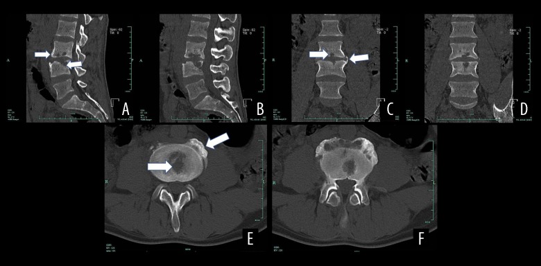
Figure 4.
CT results of a male patient aged 39 years with spinal brucellosis. Sagittal (A, B) and coronal (C, D) images revealing vertebral body lesions at the L3/4 level. The lower endplate of the L3 vertebral body and the L4 upper endplate and vertebral body exhibit bony destruction with bilateral, vertebral, bony bridge formation, as well as L3/4 endplate denaturalization and vertebral body osteogenesis (white arrows). Axial image (E, F) revealing osteolytic destruction of the vertebral body (white arrows).

