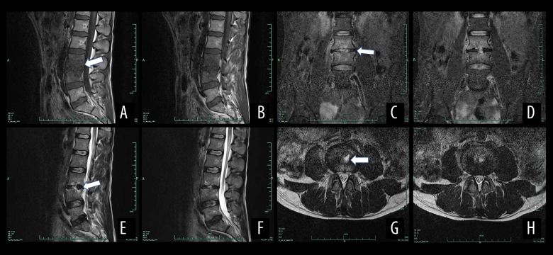
Figure 5.
Magnetic resonance imaging (MRI) results of a male patient aged 39 years with spinal brucellosis. The lesion was located at the L3/4 vertebrae. T1WI imaging of the vertebral body and low-level signaling from the intervertebral disc (A, B). On the T2WI images, the vertebral body and intervertebral disc exhibit high- and low-intensity signals, respectively (C, D), while the T2WI-FS images reveal high-intensity signals from the vertebral body and intervertebral disc (E, F). A teardrop-shaped abscess is also evident in the axial image (G, H). White arrows show the position of the lesion.

