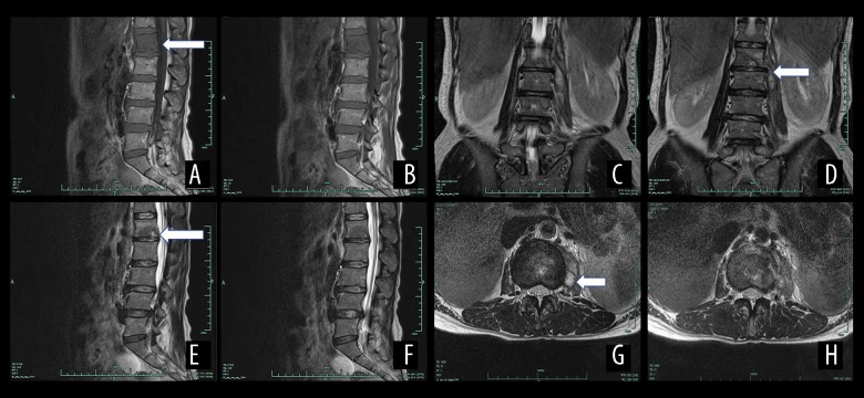
Figure 6.
The MRI results of a female patient aged 41 years with spinal brucellosis. The lesion is located at the L1/2 vertebrae. T1WI images of the vertebral body reveal low-intensity signals from the intervertebral disc (A, B). On the T2WI scans, the vertebral body and intervertebral disc exhibit high- or low-level mixed signals (C, D), and the T2WI-FS scans exhibit high-intensity signals from the vertebral body and intervertebral disc (E, F). A teardrop-shaped abscess is also evident in the axial image (G, H).

