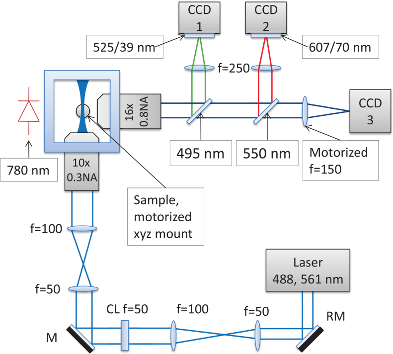
Fig. 1.
Our SPIM microscope is based around a Nikon 10× 0.3NA air objective in the launch path and a Nikon 16× 0.8NA water dipping objective in the imaging path. A Coherent Obis 488 nm laser (15 mW) was used for the tube flow experiments, and an Omicron Versalase multiwavelength laser system was used at 561 nm wavelength (30 mW) for zebrafish RBC imaging. PIV frame pairs were recorded using QImaging QIClick CCD cameras (CCD1, CCD2), and brightfield images for heart synchronization were recorded using an Allied Vision GS650 CCD camera (CCD3). Chroma T495lpxr-UF2 and T550lpxr-UF2 dichroics were used for the green and red channels respectively. Further, Thorlabs MF525-39 and Semrock FF01-607/70-25 filters were used at CCD 1 and 2 respectively. Shadow effects on the illumination arm were minimized using a resonant mirror (RM) after [22], which is essential to prevent shadow artefacts that would bias the PIV analysis.

