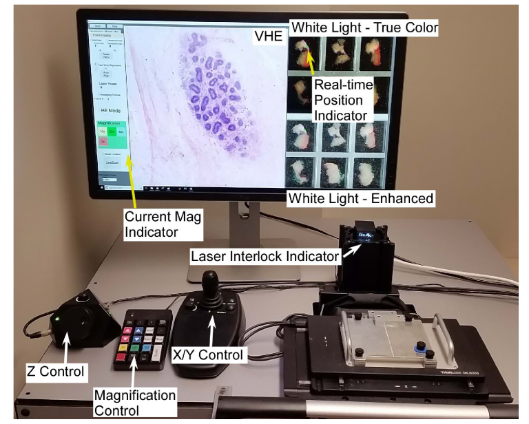
Fig. 5.
Software user interface and controls used to image freshly excised, inked bread loafed surgical specimens. The virtual H&E display shows a terminal ductal lobule unit (TDLU) imaged at 20x magnification, while the white light microscopy shows a widefield image with the current imaging location adjacent to a simulated inked margin. The user can rapidly (< 1 second) switch the magnification, while a joystick and digital focus knob are used for X/Y and Z control, respectively. An LED indicator shows the laser interlock status, enabling the operator to see when the specimen is loaded and the laser armed.

