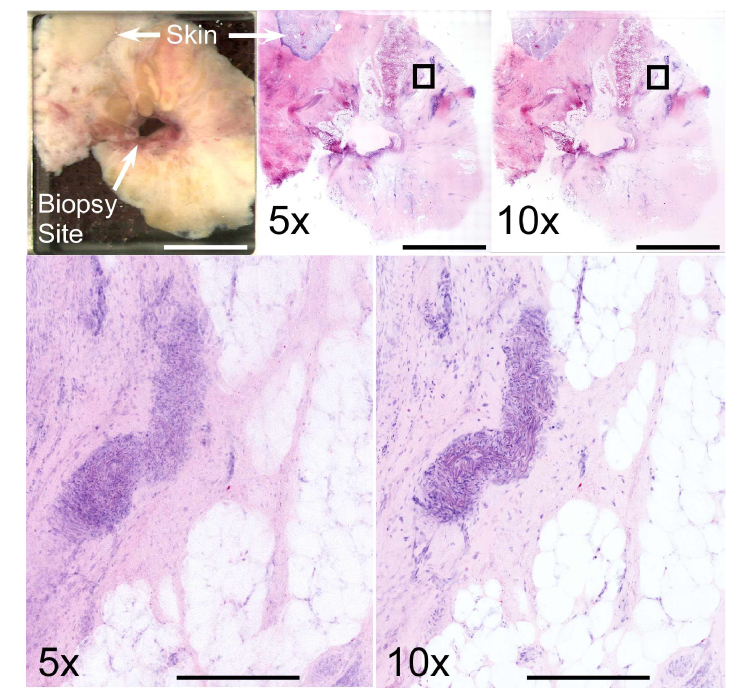
Fig. 9.
Comparison between 5x/0.25 NA and 10x/0.45 NA objectives at 1030 nm excitation wavelength using discarded superficial breast tissue excised during surgery for invasive ductal carcinoma. The biopsy site and skin are present along with large areas of fat and stroma (top left). Mosaic images viewed at low magnification show relatively limited difference between the two objectives (top center – 5x, top right – 10x), however zoomed views of the boxed regions show that cellular features are poorly resolved with the 5x/0.25 objective. In contrast, the structure of a blood vessel is readily identifiable with the 10x/0.45 objective. Scale bars: 1 cm (top) and 400 μm (bottom). Total area: 7.9 cm2. Full resolution image: http://imstore.mit.edu/system/Fig9.html.

