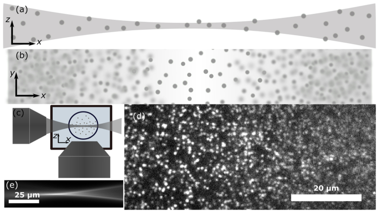
Fig. 1.
(a) The thickness of a Gaussian light-sheet varies along the direction of propagation, x. (b) Therefore, fluorescently-labeled particles may be localized at the light-sheet’s focus but not on either side where the sheet thickens. (c) Cartoon of light-sheet microscope showing the excitation objective (left) and the water-dipping imaging objective (bottom) imaging a sample within a tube placed in a water bath. (d) Image of 200-nm fluorescent beads showing good optical sectioning where the sheet is thinnest (left side) and higher background where the sheet thickens (right side). (e) Image showing the varying thickness of our light-sheet.

