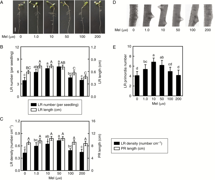
Fig. 1.
Melatonin induces lateral root (LR) formation. Three-day-old alfalfa seedlings were treated with 0, 1.0, 10, 50, 100 or 200 μm melatonin (Mel). (A) Lateral root phenotypes after treatment for 5 d. Scale bar = 1 cm. (B) Number of emerged LRs (>1 mm) per seedling and LR length. (C) Density of emerged LRs and primary root (PR) length. (D, E) After treatment for 60 h, photographs of LR primordia formation were taken and the number of LR primordia was recorded. The sample without added melatonin was the control. Means and standard errors were calculated from at least three independent experiments with at least three replicates for each. Within each set of experiments (B, C, E), bars with different letters denote significant differences (one-way ANOVA followed by Tukey’s multiple range test, P < 0.05).

