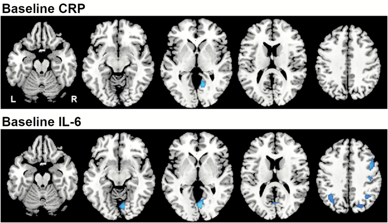
Figure 1.
Associations between baseline levels of inflammatory markers and baseline rCBF. Horizontal sections showing regions with a significant relationship between the inflammatory markers and rCBF at baseline. Blue regions represent areas of decreased activity (rCBF) in relation to higher inflammatory levels. CRP was associated with decreased rCBF in the lingual gyrus and IL-6 with decreases in the middle frontal gyrus, postcentral gyrus, lingual gyrus, superior occipital gyrus, and middle occipital cortex. Of the regions associated with IL-6, only the lingual gyrus survived correction for regional tissue volume. CRP = C-reactive protein; IL-6 = Interleukin-6; rCBF = Regional cerebral blood flow.

