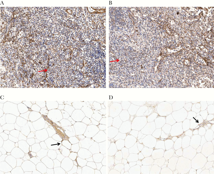
Figure 2.
Collagen I in lymph node (LN) and adipose tissue biopsy specimens. A, Substantial collagen I deposition (brown) weaving through the LN (arrow) with disruption of normal architecture. B, Low collagen I deposition in LN, with some preservation of LN architecture (arrow). C, Collagen I deposition in a vessel within adipose tissue and around adipocytes (arrow). D, Collagen I deposition in the interstitial spaces between adipocytes with inflammatory cell infiltrate (arrow).

