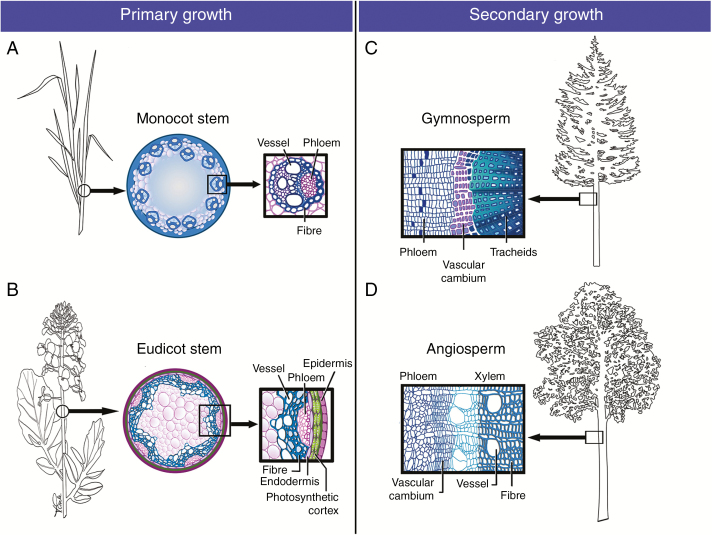
Fig. 1.
SCWs in primary and secondary stem growth. Illustrative examples of SCWs in water-conducting cells (vessels, tracheids) and supportive fibres. (A and B) SCWs in primary growth. (A) SCWs in monocot primary growth exemplified in a cross-section of a grass stem internode where vascular bundles with large metaxylem vessels are encased in SCW-rich sclerenchyma (e.g. Brachypodium). (B) SCWs in eudicot primary growth illustrated in a stem cross-section prior to onset of secondary thickening (e.g. Brassica). SCWs are found in the vascular vessels and fibres, which are continuous with the thick interfascicular fibres. (C and D) SCWs in secondary growth, marked by the presence of the vascular cambium. (C) Gymnosperm secondary growth showing thick SCWs in the water-conducting and supportive tracheids of the secondary xylem (e.g. Pinus). (D) Angiosperm secondary growth showing SCWs in the water-conducting vessels and the supportive fibres (e.g. Populus).

