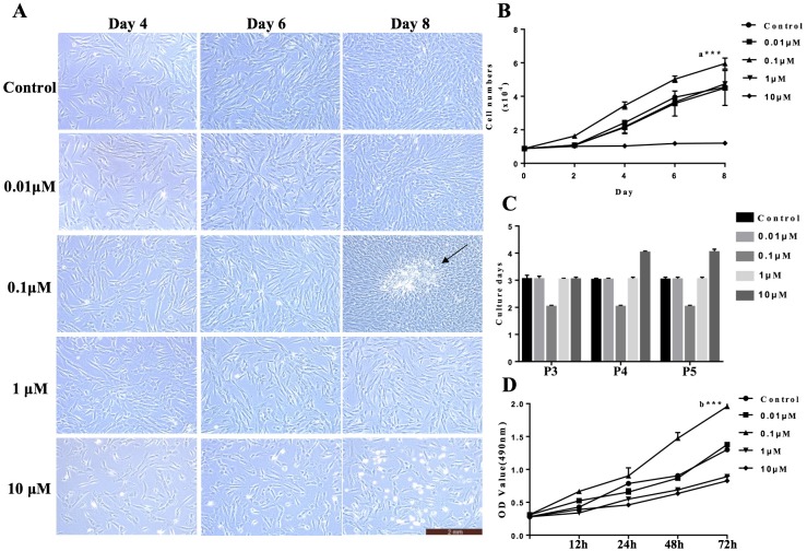
Figure 1. VB-1 facilitates the proliferation of human DPCs.
(A) Morphology of human DPCs treated with VB-1 (0–10 µM) at indicated days. Arrow indicates colony growth of DPCs. (B) Human DPCs (1 × 10⁴ cells) were plated in 24-well dishes and cultured in the presence of different concentrations of VB-1 (0–10 µM) for 8 days. Growth curves indicate the mean of three independent experiments (±SEM). (C) Culture days per passage of human DPCs treated with VB-1 (0–10 µM). Experiments were carried out in triplicates. (D) OD value of human DPCs (4 × 10³ cells) were plated in 96-well dishes and cultured in the presence of different concentrations of VB-1 (0–10 µM) for 3 days. Data are reported as mean+SEM. Student’s t-test was used to compare data. *P < 0.05, **P < 0.01.

