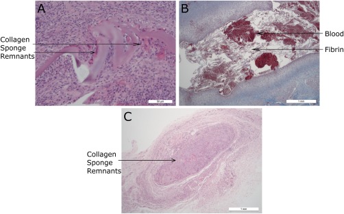
Figure 5.

Histology sections from group 2. (a) JEL #01 (HE stain) focus of granulomatous inflammation and fibrosis centered on remnants of collagen sponge. (b) JEL #02 (Masson's stain) cystic cavity filled with blood and fibrin and lined by granulomatous inflammation and granulation tissue. (c) CON (HE stain): Discrete granulomatous focus centered on residual fragments of collagen sponge.
