
Figure 4.
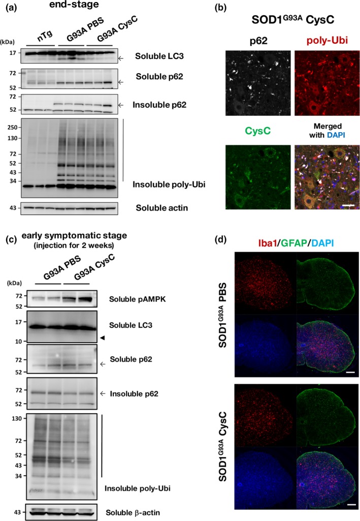
Cystatin C (CysC) administration reduces insoluble p62 and poly‐ubiquitin accumulation in the lumbar spinal cords of early symptomatic SOD1G93A mice. (a) Immunoblotting analyses of p62 and poly‐ubiquitin (poly‐Ubi). Note that detergent (1% Triton X‐100)‐insoluble p62 and poly‐Ubi was accumulated in SOD1G93A mice, but they were not affected by CysC administration. (b) Co‐localization of p62 and poly‐Ubi aggregates were predominantly observed in outside of neurons. A representative image for the lumbar spinal cord section of the end‐stage SOD1G93A mice with CysC administration stained for p62, poly‐ubiquitin, and CysC, along with a merged image with DAPI. (c) Immunoblotting analyses of spinal cords derived from the early symptomatic stage (4 months old) SOD1G93A mice administered with phosphate‐buffered saline (PBS) or CysC for 2 weeks. In contrast to the end‐stage samples (a), the amounts of insoluble p62 and poly‐ubiqutinated proteins were reduced by CysC administration, suggesting the improved protein degradation. It should be also noted that bands corresponding to LC3‐II were hardly detected (arrowhead). (d) Astrocytes and microglia in the lumbar spinal cords of end‐stage SOD1G93A mice, with or without administration of CysC, were immunostained with antibodies against glial fibrillary acidic protein (GFAP) and Iba‐1 with DAPI. Scale bars: 50 μm (b), and 100 μm (d).