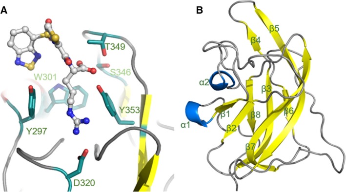
Figure 1.

Structure of the binding site of NRP1‐b1 domain. (A) Ball and stick representation of EG00229 (carbon atoms are coloured grey, nitrogen blue, sulphur yellow and oxygen red) bound to NRP‐b1 (PDB entry 3I97). NRP1‐b1 domain residues involved in the non‐covalent interactions with the ligand are shown in sticks representation. (B) Ribbon diagram of NRP1‐b1 fold: β‐sheets are represented in yellow and α‐helixes in blue.
