Figure 3.

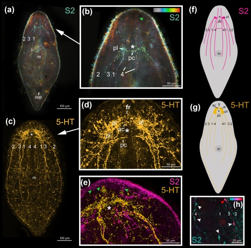
Confocal projection of Isodiametra pulchra stained with S2 antibody (a,b,e, magenta) and 5‐HT (c–e, yellow). (a) Confocal projections of a whole‐mount and (b) of the anterior body part with temportal colour coding, colour‐code: grey (dorsal) to red (ventral). Green arrowhead points to delicate anterior process of neurites. (c) Confocal projections of a whole‐mount and (d) of the anterior body‐part. (e) Overlay projection of the anterior body part of a double‐labelled animal. Green arrowheads point to delicate anterior process of neurites. (f‐g) Schematic drawings of the nervous system stained with S2 (f) and 5‐HT (g) antibodies. (h) Detailed view of bipolar and multipolar (red and white arrowheads, respectively) neurons of the colour‐coded (blue dorsal, red ventral) S2amidergic nervous system. (a,b), (c,d), (e,h) same individuals. Abbreviations: 1 dorsal neurite bundle; 2 lateral neurite bundle; 3 ventral neurite bundle; 4 medio‐ventral neurite bundle; ac: dorsal anterior commissure; al anterior lobe; cop male copulatory organ; fr frontal ring; m mouth; pc dorsal posterior commissure; pl posterior lobe. Asterisks mark position of statocyst
