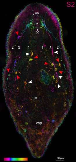
Figure 4.

Confocal projection of a whole‐mount Aphanostoma pisae stained with S2 antibody with temporal colour coding. Colour‐code: pink (ventral) to red (dorsal), arrowheads point to putative neuronal cell bodies of bipolar (white) or multipolar (red) neurons. Abbreviations: 1 dorsal neurite bundle; 2 lateral neurite bundle; 3 ventral neurite bundle; ac dorsal anterior commissure; cop male copulatory organ; fr frontal ring; m mouth; pc dorsal posterior commissure. Asterisk marks the position of the statocyst
