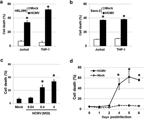
Figure 1.

HCMV‐infected cells induce cell death in surrounding cells. Jurkat and THP‐1 cells were killed following exposure to substances released from HCMV‐infected (a) HEL 299 and (b) Saos‐2 cells. (c) Death rate of Jurkat cells following exposure to HCMV‐infected HEL 299 cells. HEL 299 cells were infected with HCMV at various MOI, and co‐cultured with Jurkat cells for 72 hr. The percentage of dead cells increased with increasing viral titer in a dose‐dependent manner. (d) Death of Jurkat cells following exposure to culture supernatants isolated from HCMV‐ or mock‐infected HEL 299 cells. Culture supernatants were harvested from HCMV‐ and mock‐infected HEL 299 cells for 6 days, and incubated in the presence of Jurkat cells for 24 hr. HCMV‐infected HEL 299 cells exhibited a significantly increased death rate 2 days post‐infection. Bars indicate the means ± SEM from three independent experiments. Statistical significance between experimental means (P value) was determined using Student's t‐test.
