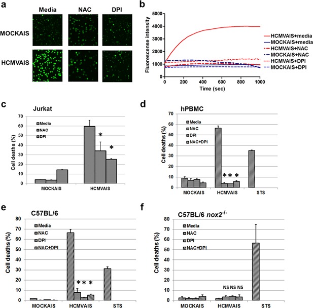
Figure 3.

NOX‐mediated production of ROS plays a role in HCMVAIS‐associated cell death. (a) ROS production by HCMVAIS. Intracellular ROS was greater in Jurkat cells following exposure to HCMVAIS (bottom left) than in MOCKAIS (top left). Intracellular ROS in Jurkat cells following exposure to HCMVAIS was significantly attenuated by treatment with NAC (bottom middle, top middle) or DPI (bottom right, top right), indicating NOX‐mediated ROS production. Representative images from live confocal microscopy (Olympus, Tokyo, Japan) at 600 s are shown; original magnification ×400. (b) The kinetics of ROS generation in live Jurkat cells treated with HCMVAIS as visualized by confocal microscopy. HCMVAIS induced intracellular ROS in Jurkat cells; no effects were evident in MOCKAIS‐treated cells. (c and d) Intracellular ROS remained at baseline amounts after treatment with the ROS inhibitors NAC or DPI: (c) Jurkat cells, (d) human PBMCs exposed to HCMVAIS. (e and f) Intracellular ROS is involved in cell death. The effects of HCMVAIS on splenocytes derived from (e) C57BL/6 wild‐type and (f) nox‐2−/− mice were examined. These results suggest that HCMVAIS‐mediated ROS production is driven by NOX. Bars indicate the means ± SEM from three independent experiments. Cell death was measured by TUNEL assay. Statistical significance between experimental means (P value) was determined using Student's t‐test. [Color figure can be viewed at http://wileyonlinelibrary.com]
