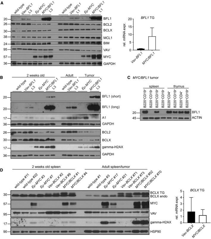
Figure 7.

MYC elevates BFL1 mRNA and protein levels (A) Spleens were dissected from 2‐week‐old wild‐type, Vav‐BFL1 (L3) TG, Eμ‐MYC TG, Eμ‐MYC/Vav‐BFL1 (L3) DT, Vav‐BFL1 (L1) TG and Eμ‐MYC/Vav‐BFL1 (L1) DT mice and, following erythrocyte depletion, cells were lysed in NP‐40 containing buffer for western analysis using antibodies specific for the indicated proteins. mRNA was isolated from spleens from 2‐week‐old Vav‐BFL1 (L3) TG and Eμ‐MYC/Vav‐BFL1 (L3) DT mice, transcribed into cDNA and assessed for BFL1 expression levels. (B) Spleens from 2‐week‐old wild‐type, Vav‐BFL1 (L3) TG, Eμ‐MYC TG and Eμ‐MYC/Vav‐BFL1 (L3) DT mice as well as from adult wild‐type and Vav‐BFL1 (L3) TG and tumour‐bearing Eμ‐MYC TG and Eμ‐MYC/Vav‐BFL1 (L3) DT mice were isolated and erythrocyte‐depleted cell preparations were lysed in NP‐40 containing lysis buffer. Western blots were probed for indicated proteins. (C) B220+CD19+ B lymphoid and B220−CD19− non‐B lymphoid cells were isolated by FACS from spleens of tumour‐bearing Eμ‐MYC/Vav‐BFL1 DT mice. Furthermore, B220+CD19+ and B220+CD19−CD4+ cells, respectively, were isolated from thymocytes of tumour‐bearing Eμ‐MYC/Vav‐BFL1 DT mice. Western blot analysis was performed as described in (A). (D) Spleens from 2‐week‐old wild‐type, Vav‐BCLX (line A) TG, Eμ‐MYC TG and Eμ‐MYC/Vav‐BCLX (A) DT mice as well as from adult wild‐type and Vav‐BCLX (A) TG and tumour‐bearing Eμ‐MYC TG and Eμ‐MYC/Vav‐BCLX (line A) DT mice were isolated and erythrocyte‐depleted cells were lysed in NP‐40 containing lysis buffer. Western blots were probed for the indicated proteins using specific antibodies. mRNA was isolated from spleens from 2‐week‐old Vav‐BCLX (line A) TG, and Eμ‐MYC/Vav‐BCLX (line A) DT mice, transcribed into cDNA and assessed for FLAG‐BCLX expression levels.
