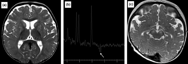
Figure 2.

(a) MRI image of Patient 2 at 9 months of age. Axial T2W image at the basal ganglia level demonstrating symmetrical hyperintense foci involving the lentiform nuclei and caudate heads. The thalami are spared. (b) MRS of Patient 2 at 9 months of age. Long TE MRS with left basal ganglia sampling demonstrating a lactate peak at 1.3 ppm. (c) MRI image of Patient 3 at 8 months of age. Axial T2W image at midbrain level, demonstrating bilateral symmetrical hyperintensity of the cerebral crura and subtle hyperintensity of the periaqueductal gray matter
