
Figure 6.
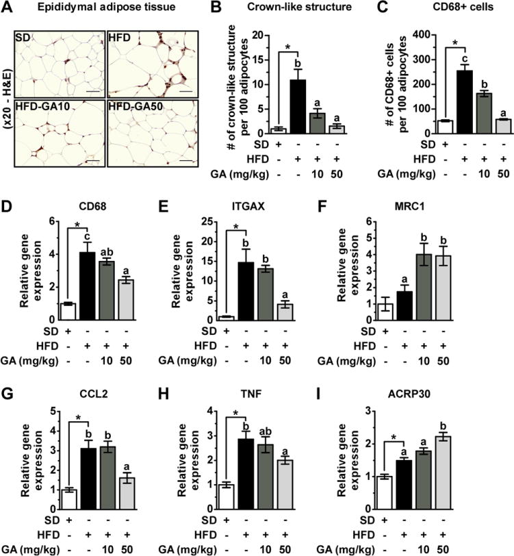
GA prevents aggravation of macrophage infiltration induced by HFD and regulates inflammation-related cytokines in EWAT. (A) Immunohisto-chemical detection of CD68 in EWAT. Macrophages are stained brown. (200×), (B) number of CLS, and (C) total number of infiltrated monocyte/macrophage into EWAT, which was quantified by counting CD68-positive cells per 100 adipocytes. (D) CD68, (E) ITGAX, and (F) MRC1 genes expression in EWAT. (G) CCL2 (H) TNF, and (I) ACRP30 genes expression in EWAT. All values were normalized to GAPDH.