Figure 3.
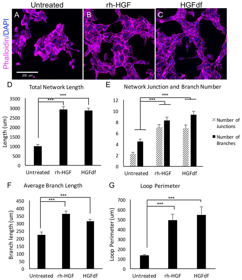
In vitro angiogenesis in response to HGFdf treatment. Representative micrographs of human umbilical cord endothelial cells cultured in basal media and then (A) left untreated, treated with (B) rh-HGF or (C) HGFdf. Treatment with rh-HGF or HGFdf leads to (D) a significant increase in total network length, (E) a significant increase in the number of junctions and branches, (F) a significant increase in branch length, and finally, (G) increased loop perimeter (***p<0.001).

