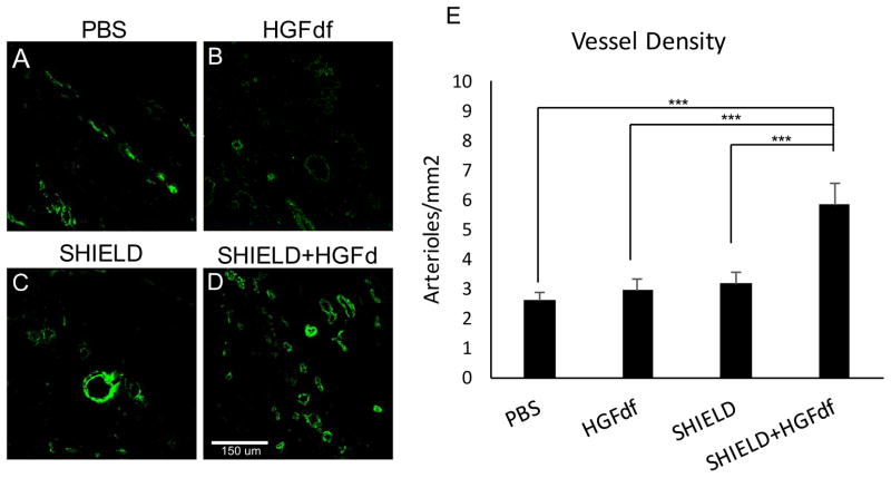
Figure 7.
Histological characterization and quantification of arterial density (stained for alpha- SMA). Representative confocal images are shown for explanted MI tissue treated with (A) PBS, (B) HGFdf, (C) SHIELD, and (D) SHIELD+HGFdf. (E) Analysis of immunofluorescence expression of α-SMA revealed a significant increase in arteriole formation in the SHIELD+HGFdf group compared to all other study groups (***p<0.001).

