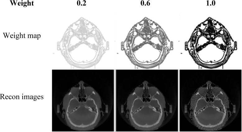
Figure 12.

Comparisons of clinical head images reconstructed via PCTV using different weighting factors using 50 half-fan projections. Weighting factors are increased from 0.2, 0.6 to 1.0 from left to right columns. First row is weight map while the second row is reconstructed images with corresponding upper map.
