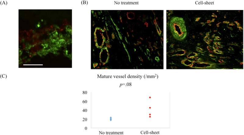
Figure 2.

(A) The bi-level cell sheet maintained alpha smooth muscle actin (SMA) positive smooth muscle cells (SMCs) and CD31 positive endothelial progenitor cells (EPCs) in separate layers in vitro. Green indicates alpha SMA; red, CD31. White bar = 100 um. (B) Representative images demonstrating von Willebrand factor (vWF) and alpha smooth muscle actin (SMA) staining of border zone myocardium for cell sheet-treated and untreated groups. Green indicates alpha SMA; red, vWF. (C) Quantification of arterial density. Mature artery density was increased in the cell sheet-treated group compared with the untreated group.
