. 2015 Jul 22;6(10):5409–5418. doi: 10.1039/c5sc01955a

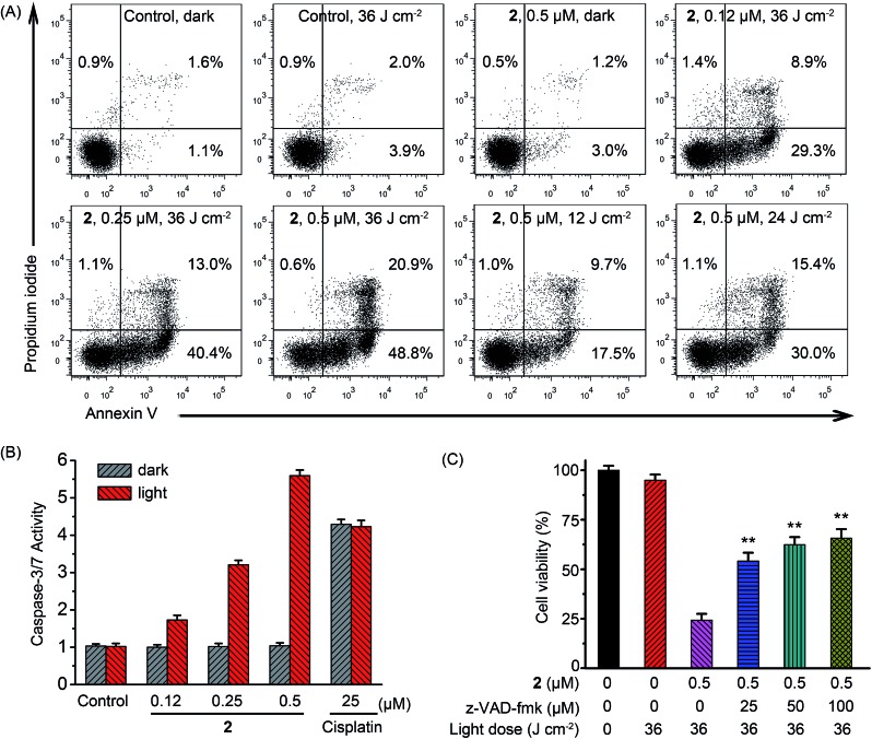
Fig. 7. (A) Flow cytometric quantification of annexin V and propidium iodide double-labeled A549 cells treated with 2 for 24 h in the absence or presence of light at the indicated concentrations. After A549 cells were incubated with vehicle or 2 for 12 h, the cells were irradiated with a 425 nm LED light for 15 min (36 J cm–2). (B) Detection of caspase-3/7 activity in A549 cells after treated with 2 or cisplatin in the absence or presence of light at the indicated concentrations. Dark: cells were incubated with 2 or cisplatin for 24 h in the dark. Light: cells were incubated with 2 or cisplatin for 12 h in the dark and then irradiated with light at 425 nm (36 J cm–2) and further incubated for 12 h in the dark. (C) z-VAD-fmk concentration-dependent inhibition against A549 cell death induced by 2-mediated PDT. Data are represented as means ± SD of three independent experiments. **P < 0.01, as compared with the group treated with 2 (0.5 μM) in the absence of z-VAD-fmk.

Fig. 7