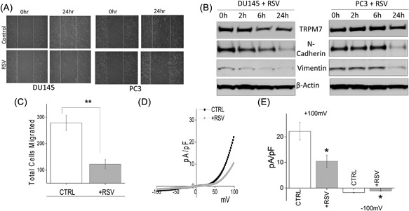
Figure 4.

(A) Wound healing assay images for cell migration in human prostate cancer DU145 and PC3 cell lines pretreated for 24 h with 50 μM RSV. (B) Western blot showing the expression of vimentin, N‐cadherin, TRPM7, and loading control β‐actin in DU145 and PC3 cells with RSV treatment under different time points. (C) DU145 cells were migrated using transwell inserts and serum free RPMI ± 50 μM RSV. Four random fields at 10× were counted from seven separate experiments indicating total cells migrated ± SEM. ** indicate significance (P < 0.01). (D) IV curves of TRPM7 current under various conditions (control or RSV pretreatment) and quantitation (5‐10 recordings) of current intensity at ±100 mV is shown in (E). * indicate significance (P < 0.05)
