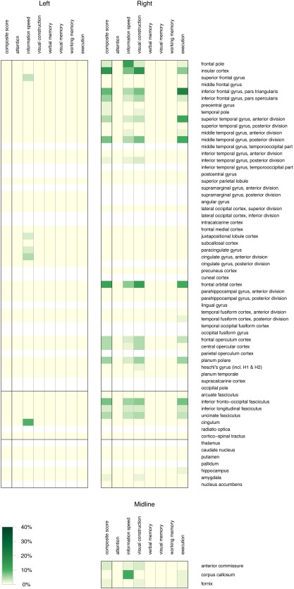
Figure 4.

Heat map of percentage of voxels (shades of green) with a q value <0.4 in a cognitive domain in columns per anatomical region in rows. The anatomical regions are grouped by laterality and midline structures, and subdivided by horizontal black bars in cortical regions, subcortical white matter pathways, and subcortical grey nuclei. No brain regions were identified for decline in attention, verbal memory, visual memory, and working memory, and therefore, these rows are empty. Absence of information in anatomical regions is demonstrated as white, whereas information of 0% is demonstrated as light yellow [Color figure can be viewed at http://www.wileyonlinelibrary.com]
