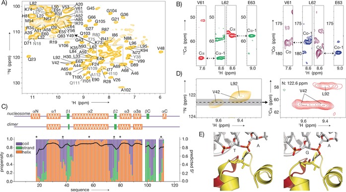
Figure 2.

Resonance assignment and secondary structure of H2A in sedimented nucleosomes. A) 2D NH spectrum with (tentative) assignments indicated in (gray) black. Side chain resonances in light colors. B) Representative strips illustrating the sequential backbone assignment based on 3D CANH and CA(CO)NH (red and green) or CONH and CO(CA)NH (magenta and blue) spectra. C) Secondary structure propensities (colored bars) and predicted S2 values (black line) based on assigned backbone chemical shifts. Secondary structure in the nucleosome crystal structure (PDB ID 2PYO18) and isolated H2A–H2B dimer (unpublished results) shown at the top. Asterisks indicate tentative assignments. D, E) Peak doubling of V42 observed in the 2D NH (left) and 3D CANH (right) spectra (D), correlated to the asymmetric environment in 601 nucleosomes (PDB UD 3LZ020). Dashed lines indicate hydrogen bonds (E).
