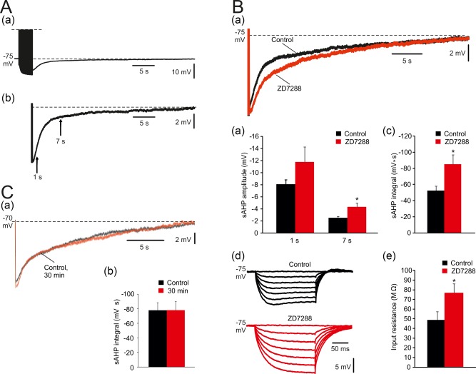
Figure 1.

Prolonged sAHPs evoked by spike trains in CA1 pyramidal cells. (A. a) In a representative neuron, sAHPs were evoked by a 50 Hz train of 150 spikes lasting about 35 s. Each spike was evoked by a brief suprathreshold depolarizing current pulse. Here and below, each sAHP trace is the average of three consecutive recordings obtained at 3‐min intervals. (b) Enlarged trace of a sAHP depicting the time points for measuring early (1 s) and late (7 s) sAHP amplitudes (indicated by arrows). Dashed lines represent baseline Vm, used to delineate “area under the curve” representing sAHP integral. (B) Effects of blocking HCN channels on sAHPs evoked by 150 stimuli. (a) Representative traces; application of 50 μM ZD7288 caused an increase in sAHP amplitude and integral. The duration of the sAHP was not affected. (b and c) Bar diagrams depicting the pooled results (mean ± SEM; n = 8) for sAHP amplitudes and integrals, respectively. Note that only sAHP amplitude at 7 s post‐stimulus and sAHP integral were significantly increased by ZD7288. (d) Representative traces; in the same 8 neurons, application of ZD7288 caused an increase in RN and abolished the “sag” of Vm during the hyperpolarizing responses to the negative current pulses. (e) Bar diagram showing the significant RN increase in ZD7288 (mean ± SEM). (C. a) representative traces; the sAHPs evoked in CA1 pyramidal cells by trains of 150 spikes were stable for 30 min. Here and below, unless otherwise stated, all normal and modified aCSFs contained 50 μM ZD7288. (b) Bar diagram depicting the pooled (mean ± SEM; n = 10) and showing the stability of sAHPs over periods of 30 min [Color figure can be viewed at http://wileyonlinelibrary.com]
