Figure 2.

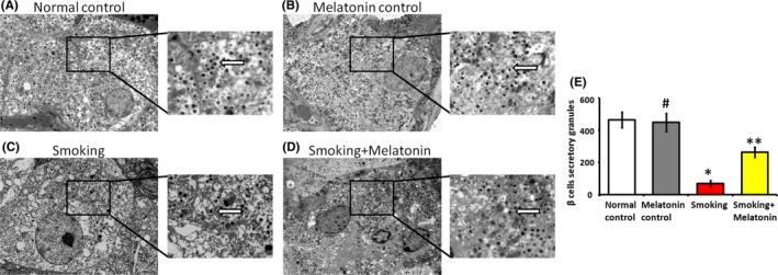
The secretory granules (white arrow) and cytoarchitecture of β cells were examined by electron microscopy. (A) The β cells of normal control rats: abundant secretory granules and a clear cellular structure are evident. (B) The β cells of melatonin control rats: abundant secretory granules and a clear cellular structure are evident. (C) The β cells of smoking rats: the secretory granules are sparse, and the faint cytoarchitecture can be observed. (D) The β cells of smoking rats with intraperitoneally injecting melatonin: compared with the β cells of smoking rats, increased number of secretory granules and distinct cytoarchitecture can be observed. (E) The column diagram shows the number of secretory granules in the different groups. 20 000× magnification. #P > .05 melatonin control vs normal control, *P < .05 smoking vs normal control, **P < .05 smoking + melatonin vs smoking
