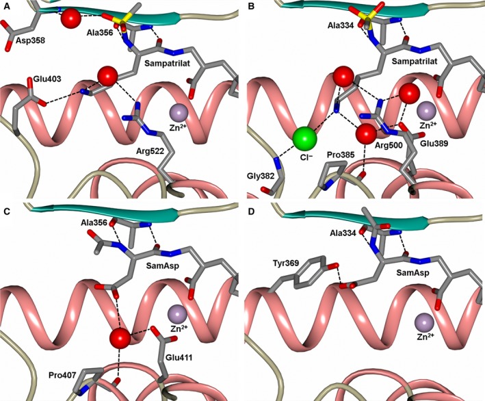
Figure 7.

Close up views of (A) sampatrilat‐cACE, (B) sampatrilat‐nACE, (C) samAsp‐cACE and (D) samAsp‐nACE binding sites showing the H‐bond/electrostatic interactions in the S1 and S2 subsites. The protein chain is shown as a cartoon with α‐helices and β‐strands in rose and dark cyan, respectively, and water molecules are depicted as red spheres.
