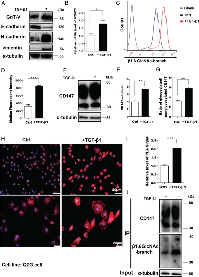
Figure 1.

The level of CD147‐β1,6‐GlcNAc‐branched structures is enhanced during EMT. (A) Cells were treated with 2.5 ng/ml TGF‐β1 for 24 h and cell lysates were analyzed by western blotting. (B) Real‐time PCR detection of MGAT5 mRNA levels in QZG cells stimulated with 2.5 ng/ml TGF‐β1 for 24 h. GAPDH was used as the normalization control. (C, D) Membrane levels of β1,6‐GlcNAc branching were measured by FACS with fluorescein‐labeled PHA‐L in QZG cells stimulated with TGF‐β1 for 24 h. (E–G) Western blotting of CD147, CD147/tubulin, and glycosylated/non‐glycosylated CD147 in control and TGF‐β1‐treated cells. (H, I) In situ PLA in control cells and cells treated with TGF‐β1. (H) Representative image. (I) Quantification. (J) QZG cells treated with TGF‐β1 were subjected to CD147/HAb18G immunoprecipitation (IP), western blotting, and lectin analyses. The results are shown as the mean ± SD (n = 3). Significance was determined by Student's t‐test (*p < 0.05; **p < 0.01; ***p < 0.001).
