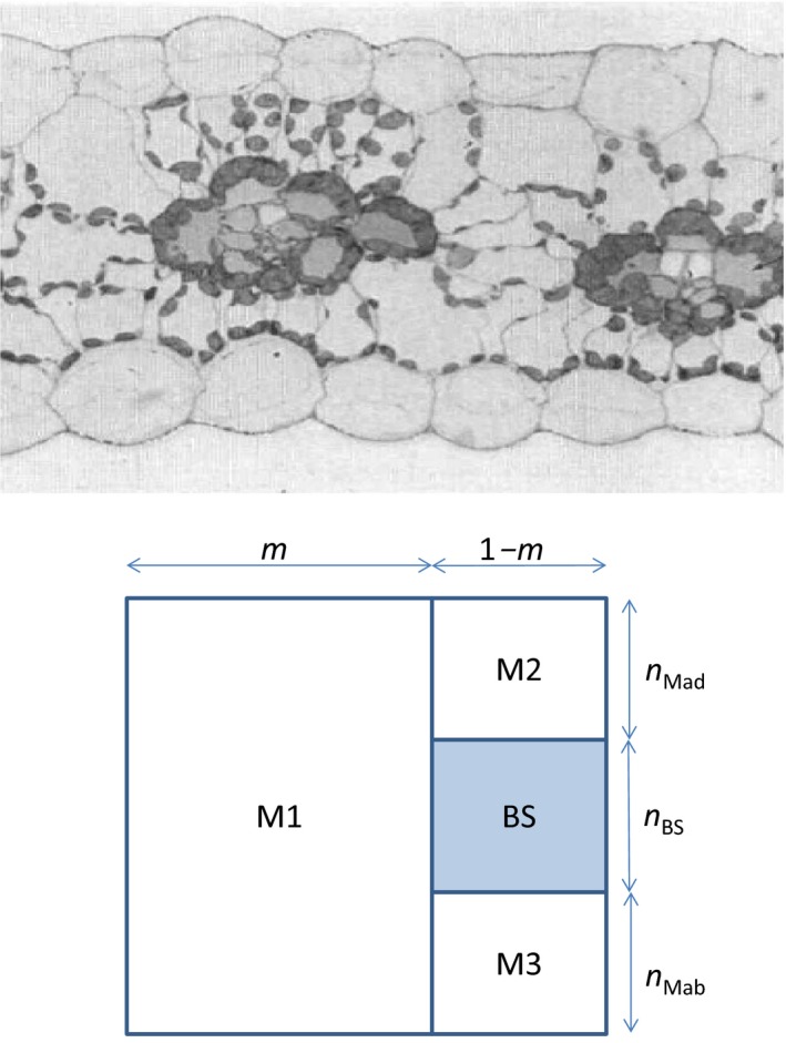
Figure 1.

Upper panel: two units of interveinal distance of a maize leaf (redrawn from Evans & von Caemmerer, 1996; with permission); lower panel: schematic representation of bundle‐sheath (BS) and mesophyll (M) sections of one unit of interveinal distance. The BS section is shaded, with relative height n BS. The M part has three sections: interveinal M (M1), the adaxial side above BS (M2) and the abaxial side below BS (M3). The relative heights of M2 and M3 are denoted as n Mad and n Mab, respectively; thus, the relative height of M1 is (n Mad + n BS + n Mab), which together makes one full relative height. In the model, it is assumed for simplicity that n Mad = n Mab = (1‐ n BS)/2, based on many published images of C4 leaves (e.g. Ghannoum et al., 2005). The fraction of one unit interveinal distance for M1 is m; the remaining fraction, 1‐ m, is then the vein width. So, areas of BS, M1, M2 and M3 sections can be easily calculated from m and n BS. The total Chl content in M cells can be partitioned among the three M sections according to their areas relative to the total M area. In a real leaf, M1 is commonly divided into two pieces that are placed on both left and right sides of the M2–BS–M3 vein area (Bellasio & Lundgren, 2016), and the two subsections of M1 and the M2–BS–M3 section together form the interveinal distance. Here, the two subsections are combined into a single M1 for mathematical simplicity, but this has no influence on our modelling results.
