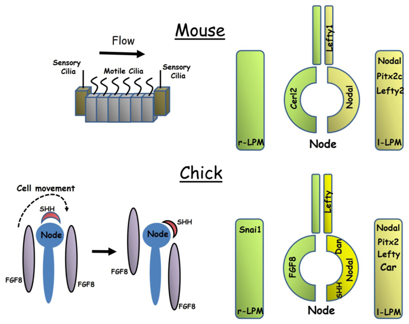
Figure 1.
The mouse and chick embryos represent the two major mechanisms of symmetry breaking seen in vertebrates. In the mouse (top left), a leftward fluid flow is generated across the node by rotational beating of motile cilia while in the chick embryo (lower left) a leftward movement of cells transforms the initially symmetrical expression of Shh and Fgf8 into an asymmetric one. The diagrams on the right illustrate similarities and differences between the two embryos in the expression of key genes within the node, lateral plate mesoderm (LPM) and midline floorplate. Note that many additional genes not shown are also asymmetrically expressed in chick.

