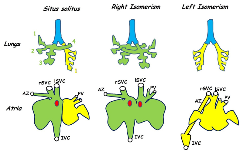
Figure 3.
Isomerism is seen in both the lungs (top row) and in the atria (bottom row). This figure shows the anatomy of the mouse. Situs solitus is the normal morphology in which the right lung has four lobes (green) while the left has only one (yellow). Animals with left-right patterning defects may develop symmetrical lung morphology with either four lobes on each side (right isomerism) or one on each side (left isomerism). The atria differ both in morphology of the appendages (exaggerated in the figure for diagrammatic purposes) and in venous connections. In normal hearts both superior venae cavae (rSVC, lSVC) as well as the inferior vena cava (IVC) enter the right atrium while the pulmonary veins (PV) enter the left (situs solitus). The azygous vein (AZ) drains into the right superior vena cava. In hearts showing right isomerism the right SVC (rSVC) enters the right side while the left (lSVC) enters the left. The sino-atrial node (SAN, red dot) is duplicated and there is an atrial septal defect. Left isomerism is also associated with an atrial septal defect and both the venae cavae and pulmonary veins enter near the middle of a common atrium while the inferior vena cava is interrupted and drains into one of the superior venae cavae via the azygous vein. Variations are seen between the mouse and human phenotype (see text). Adapted from [63,67,68].

