Figure 5.
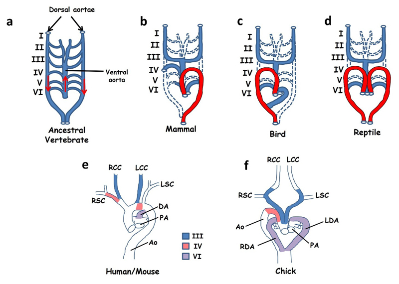
(a–d) Simplified cartoons to illustrate remodelling of the arterial circulation during embryogenesis. Ancestral vertebrates and modern day fish (a) use a symmetrical arterial system in which blood leaves the heart through a single ascending ventral aorta before entering a series of six paired arches (blood flow is indicated by red arrows). These vessels feed into a pair of descending dorsal aortae that carry oxygenated blood to the viscera. A similar circulation system is seen in early-mid stage embryos of all vertebrates. Remodelling during late embryogenesis involves regression of some vessels (dashed lines) with retention and expansion of others. Different remodelling strategies lead to a leftward looped aortic arch (shown in red) in mammals (b), a right arch in birds (c) and a double aorta in reptiles (d). Images in (e) and (f) show the anatomy of the great vessels in mice and humans at the time of birth (e) and in the chick at the time of hatching (f). The contributions of pharyngeal arch arteries III, IV and VI are indicated. (a–d) Adapted from [98] (e–f) adapted from [99]. Abbreviations: Ao: aorta, DA: ductus arteriosus, LCC: left common carotid, LDA: left ductus arteriosus, LSC: left subclavian, RCC: right common carotid, RDA: right ductus arteriosus, RSC: right subclavian.

