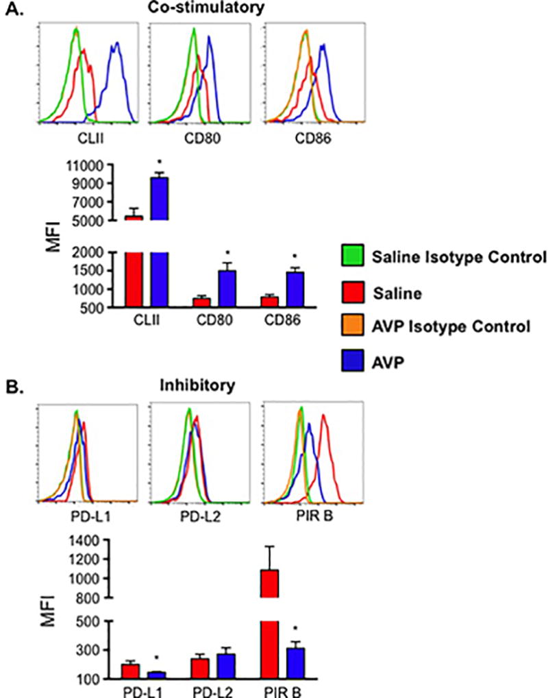
Figure 4.

AVP induces altered surface receptor expression on DC. Inhibitory molecules paired immunoglobulin-like receptor B (PIR B) and programmed death ligand 1 (PD-L1) are decreased while co-stimulatory molecules MHC Class II (CLII), CD80, and CD86 were increased on dendritic cells from AVP infused dams. Lymphocytes were isolated from the spleen of saline and AVP infused dams. Following fluorescent antibody staining, CD11c+ DCs were gated as shown in Supplemental Figure 1B. (A) Representative histograms and mean fluorescence intensity (MFI) of co-stimulatory cell surface molecules. (B) Representative histograms and MFI of inhibitory cell surface molecules. Background fluorescence was determined by staining cells with corresponding fluorochrome-conjugated rat immunoglobulin isotype control antibodies. Isotype control subtracted MFIs are shown. N ≥ 5 per group from at least two independent experiments. MFIs are mean ±SEM. Statistical significance was determined using a Student t test and the minimal level of confidence deemed statistically significant was a p value <0.05. *=p<0.05.
