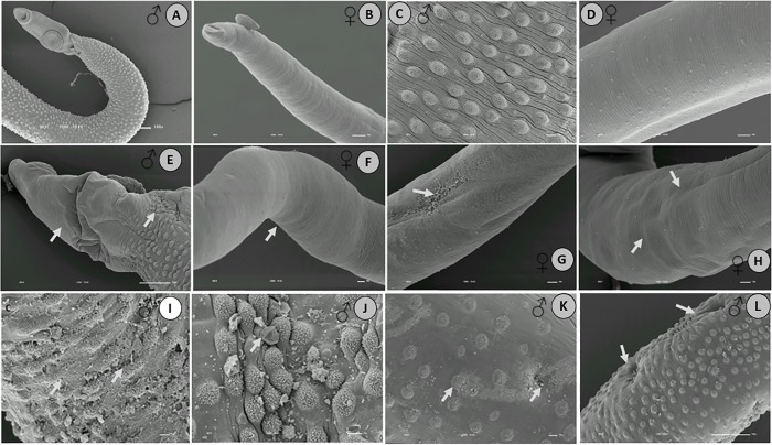
Fig 6. Scanning electron microscopy of S. mansoni adults retrieved 48 h after treatment with EPIIS (100 mg/kg).
A-D: Control group—untreated; E–L: Male and female worms. The arrows show damage to tegument with reduction in the size of the spines, and damage to the oral and ventral suckers in male worms, followed by body corrugation in female worms. Image bar scale: (A) 100X___10 kv; (B) 120X ___10 kv; (C-D) 180X ___10 kv; (E-F) 127X ___10 kv; (G-H) 250X___10 kv; (I- L) 350X___10 kv.

