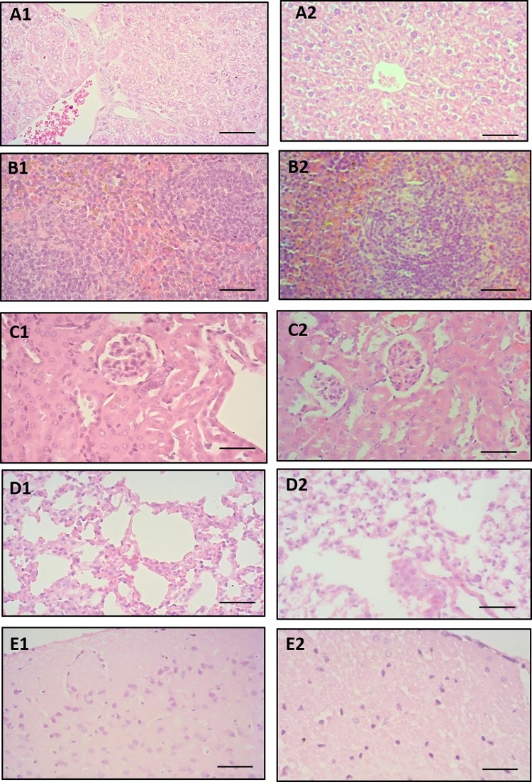
Fig 9.
Photomicrographs of histological sections of liver (A1, A2), spleen (B1, B2), kidney (C1, C2), lung (D1, D2) and brain (E1, E2) obtained from Swiss mice of control group (first column) and group treated with 2000 mg/kg EPIIS (second column). Hematoxylin & eosin, 400 X magnification, bars = 50 μm.

