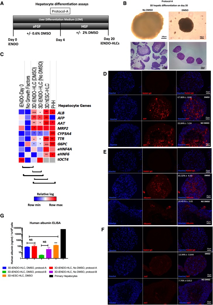
Fig 4. 14TF iENDO cells differentiate into hepatocyte like cells.
A) Protocol time-line for hepatocyte differentiation in 3D from iENDO cells protocol A with or without DMSO. B) Morphology of iENDO differentiated organoids at day 20 with and without DMSO by bright field and H&E staining (N = 3). C) Relative gene expression (to PPIG, log scale) in day 20 iENDO-HLCs ± DMSO represented as a heat-map for hepatocyte markers compared with hESC-HLCs organoids (d30) and PHHs (N = 3). D-F) Immunostaining for AFP, ALB and AAT on day 20 iENDO organoids ± DMSO with respective isotype controls. Scale bar 25 μm (representative example of N = 3). G) Albumin secretion in supernatants of day 20 iENDO organoids ± DMSO, hESC day 30 organoids and primary hepatocytes (PHH) (N = 3). Error bars represents standard deviation of three independent experiments. *p<0.05, **p<0.01 and ***p<0.001 determined by unpaired 2-tailed Student’s t-test. NS- Not significant.

