
Figure 1. OCT protein expression in UC-MSCs.
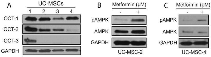
(A) Whole cell lysates extracted from commercially available, human-derived UC-MSCs obtained from four different donors were analyzed by Western blotting to determine the expression levels of OCT-1, OCT-2 and OCT-3. Whole cell lysates obtained from UC-MSC-2 (B) and UC-MSC-4 (C) following a 3-hour treatment with metformin (10 μM) demonstrate an increase in the phosphorylating status of AMPKα1 at Thr172 (pAMPK) as analyzed by Western blotting. In all immunoblots GAPDH served as loading control.