
Figure 4. Metformin significantly increases mineralized nodule formation in UC-MSCs.
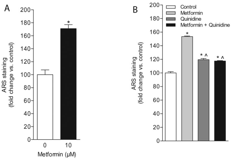
(A) Quantification of ARS-stained, calcium-containing nodules in UC-MSC-4 cells in response to metformin following a 21-day treatment. Data represent mean ± S.E.M. *p<0.05, when compared to untreated control. (B) Treatment of UC-MSC-4 cells with metformin (10 μM) and quinidine (10 μM) for 21 days resulted in a significant decrease in the formation of calcium-rich nodules as determined by ARS staining. Data represent mean ± S.E.M. *p<0.05, when compared to untreated control; ˆp<0.05, when compared to metformin-treated cells.