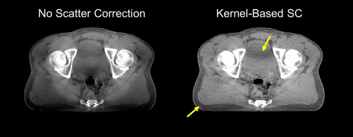
Figure 1.

Reconstructed cone‐beam CT images of a clinical pelvis scan. Severe shading is seen when no scatter correction is applied. Even after kernel‐based (fASKS) scatter correction, some residual scatter artifact remains, including shading in the bladder (arrows). Display window [−300, 300] HU. [Color figure can be viewed at wileyonlinelibrary.com]
